Intended for US audiences only.

Top 3 Tests Used to Diagnose Primary Immunodeficiency

Detecting primary immunodeficiency (PI) can be challenging because it typically manifests through a variety of chronic illnesses. Once PI is suspected as the underlying cause, there are three main ways to test for and diagnose it: laboratory testing, newborn screening, and genetic inheritance / familial history. You can learn about the three main diagnosis methods here:

Laboratory
Testing
Newborn
Screening
Genetic
Inheritance

Laboratory Testing

Laboratory testing plays a central role in the evaluation of the immune system and identification of PI and its specific type. An accurate medical history, family history and physical examination are critical to developing the best strategy for laboratory evaluation. This typically begins with screening tests, followed by more sophisticated (and costly) tests chosen based on the initial test results.

Lab tests evaluate several values, looking to see which fall within or outside of what is considered “normal range”, based on a statistical analysis of similar populations. While not exact pointers — finding a value outside of the reference range does not automatically represent an abnormality — abnormal laboratory values signal signs of PI disorders: antibody deficiencies, cellular (T-cell) defects, neutrophil disorders, or complement deficiencies.

Laboratory Evaluation for Antibody Deficiency, or Humoral Immunity

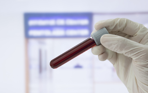
The standard screening test for antibody deficiency starts with measurement of immunoglobulin (Ig) levels in the blood serum, evaluated through testing of blood samples. These test for IgG, IgA and IgM levels. There are other tests for specific antibody production. These tests measure how well the immune system responds to vaccines. Additional studies used to evaluate patients with antibody deficiencies include measuring the different types of lymphocytes in the blood by marking those cells with molecules that can identify the different types. In addition, analysis of DNA can be used to confirm a particular diagnosis. Finally, there are studies done in specialized laboratories to assess immunoglobulin production by cultured lymphocytes in response to a variety of different kinds of stimuli.

An antibody is a blood protein produced to fight substances that the body recognizes as foreign, such as a bacteria or a virus.


Evaluation of Cellular (T-Cell) Immunity

The laboratory evaluation of cellular or T-cell immunity focuses on determining the numbers of different types of T-cells and evaluating the function of these cells. The simplest test to evaluate possible decreased or absent T-cells is a complete blood count (CBC) and differential to establish the total blood (absolute) lymphocyte count. The measurement of the number of T-cells is often accompanied by cell culture studies that evaluate T-cell function. This is done by measuring the ability of the T-cells to respond to different types of stimuli. There are an increasing variety of functional tests that are available to evaluate T-cells. An immunologist is the best person to undertake this interpretation. Many immune deficiencies are associated with specific genetic defects. This is particularly true of Severe Combined Immune Deficiency (SCID) where more than 12 different genetic causes for SCID have been identified. These can all be evaluated using current technology for mutation analysis, and this is the most accurate means to establish the definitive diagnosis.

T-cells are a specific sub-type of white blood cell, a lymphocyte, which send out chemical instructions to the body on how to best fight bacteria, viruses, or parasites.


Evaluation of Neutrophil Function

The laboratory evaluation of the neutrophil begins by obtaining a series of white blood cell counts (WBC) with differentials. The WBC and differential will determine if there is a decline in the absolute neutrophil count, a condition called neutropenia. This is the most common abnormal laboratory finding when a patient presents with a clinical history that suggests defective neutrophil immunity. Usually more than a single CBC and differential is necessary to diagnose neutrophil problems. Further tests of neutrophils — measurement of the creation of reactive oxidation or cytometry to assess the presence of certain proteins — are then made to determine the particular type of PI disease that may be present.

A neutrophil is a specific type of white blood cell key to the identification of, and initial fight against, infection.


It is the direct link between the clinical findings and laboratory testing that has extended our understanding of PI diseases. The continuation of this trend and laboratory testing of the future will likely be even more sophisticated and help provide further answers to the underlying basis of the expanding range of PI disease.

Back to top

Newborn Screening

Severe Combined Immune Deficiency (SCID – pronounced “skid”) leads to life-threatening infections unless the immune system can be restored through a bone marrow transplant, enzyme replacement or gene therapy. Infants with SCID who lack a family history have been diagnosed in the past only after developing serious infections. Early identification of SCID through screening of all newborns can make possible life-saving intervention before infections occur.

Currently, several states have adopted the T-cell receptor excision circle (TREC) assay as part of their routine newborn screening programs. TREC screening has identified infants with most forms of SCID and also some infants with very low T-lymphocytes due to other conditions, facilitating early, preventive medical intervention. The goal is to make TREC part of every state’s newborn screening panel.

Screening Test for SCID: T-cell Receptor Excision Circles

T-cell receptor excision circles (TREC) are circular DNA molecules formed within T-cells developing in the thymus. TREC DNA circles are measured in the blood by a technique called polymerase chain reaction (PCR). Normal infant blood samples have one TREC per 10 T-cells, reflecting the high rate of new T-cell generation early in life. Infants with SCID lack TREC altogether.

T-cells are a specific sub-type of white blood cell, a lymphocyte, which send out chemical instructions to the body on how to best fight bacteria, viruses, or parasites.


Why Screen for SCID?

The absence of T-cell and antibody immunity causes severe infections, diarrhea and failure to thrive. These are the problems that bring infants with SCID to medical attention. The infections experienced by the child with SCID are often caused by opportunistic organisms, ones that would not make a child with an intact immune system ill. Prior to 1968, when the first successful bone marrow transplant was performed, SCID was always fatal. Now it can be treated by transplantation of bone marrow stem cells from a healthy donor, or by enzyme replacement or even gene therapy.

Knowing how SCID is inherited has permitted some families, often following tragic loss of an affected infant due to infection, to make the diagnosis in subsequent affected children at birth, or even before birth. In these circumstances, early treatment of infants with SCID who have avoided infections has led to a very high likelihood of survival free of complications. Population-based newborn screening for SCID is based on the recognition that pre-symptomatic identification and treatment would improve survival for all infants born with SCID, not just those with a known affected relative.

Back to top

Genetic Inheritance / Familial History

Many diseases are genetic in origin and are passed on in families. Most of the PI diseases are inherited in one of two different modes of inheritance: X-linked recessive or autosomal recessive; rarely, the inheritance is autosomal dominant. The different modes of inheritance are associated with particular types of PI disease and who develops the symptoms of that disease.

Laboratory studies can be helpful in establishing the possible role of genes or chromosomes in a particular PI disease. In addition, family history information may help to identify a particular pattern of inheritance, as can comparisons to other families with similar problems.

Types of Inheritance

X-linked Recessive Inheritance

In X-linked recessive inheritance the PI disorder only affects males.

Since women have two X chromosomes, they usually do not have problems when a gene on one X chromosome does not work properly. This is because the second X chromosome usually carries a normal gene and compensates for the abnormal gene on the affected X chromosome. Men have only one X chromosome, which is paired with their male-determining Y chromosome. The Y chromosome does not carry much active genetic information. Therefore, if there is an abnormal gene on the X chromosome, the paired Y chromosome has no normal gene to compensate for the abnormal gene on the affected X chromosome, and the boy (man) will have the disorder. This special type of inheritance is called X-linked recessive.

In this type of inheritance, a family history of several affected males may be found. The disease is passed on from females (mothers) to males (sons). While the males are affected with the disease, the carrier females are generally asymptomatic and healthy even though they carry the gene for the disease because they carry a normal gene on the other X chromosome.


Examples of PI Diseases with X-Linked Recessive Inheritance:

  • X-Linked Agammaglobulinemia (XLA)
  • Wiskott-Aldrich Syndrome
  • Severe Combined Immune Deficiency (SCID), caused by mutations in the common gamma chain
  • Hyper IgM Syndrome, due to mutations in CD40 ligand
  • X-Linked Lymphoproliferative Disease, two forms
  • Chronic Granulomatous Disease (CGD), the most common form

The chances for a given egg combining with a given sperm are completely random. According to the laws of probability, the chance for any given pregnancy of a carrier female to result in each of these outcomes is as follows:

  • Carrier female: 1 in 4 chance or 25%
  • PI-symptomatic male: 1 in 4 chance or 25%
  • Normal female: 1 in 4 chance or 25%
  • Normal male: 1 in 4 chance or 25%

Autosomal Recessive Inheritance

In automosomal recessive inheritance the PI disorder can affect both males and females.

If a PI disease can only occur if two abnormal genes (one from each parent) are present in the offspring, then the disorder is inherited as an autosomal recessive disorder. If an individual inherits only one gene for the disorder, then he or she carries the gene for the disorder but does not have the disorder itself.

In this form of inheritance, males and females are affected with equal frequency. Both parents carry the gene for the disease although they themselves are healthy.

Examples of Autosomal Recessive Inheritance:

  • Severe Combined Immune Deficiency, several forms
  • Chronic Granulomatous Disease, several forms
  • Ataxia-Telangiectasia

The chances for a given egg to combine with a given sperm are completely random. According to the laws of probability, the chance for any pregnancy of carrier parents to result in each of the following outcomes is as follows:

  • Affected child: 1 in 4 chance or 25%
  • Carrier child: 2 in 4 chance or 50%
  • Normal child: 1 in 4 chance or 25%

Autosomal Dominant Inheritance

In automosomal dominant inheritance the PI disorder can affect both males and females.

In rare situations, a normal gene in the presence of a mutated gene cannot compensate for the defective gene; in this situation, the abnormal gene is said to exert a “dominant negative effect.”

Examples of Autosomal Dominant Inheritance:

  • Hyper IgE Syndrome, due to mutations in STAT3 (Jobs syndrome)
  • Warts, Hypogammaglobulinemia, Infections and “Myelokathexis” (a form of neutropenia – low neutrophil counts) (WHIM syndrome)
  • DiGeorge Syndrome
  • Some rare forms of defects in the IFN-γ/IL-12 pathway

Because the chances that a given egg combines with a given sperm (normal or with the HIES gene mutation) are completely random, the chance to have an affected child in this situation is 50% (2 in 4 possible outcomes).


Carrier Testing

In many primary immunodeficiency diseases, carrier parents can be identified by laboratory tests. Consult with your physician or genetic counselor to learn if carrier detection is available in your specific situation.


Reproductive Options

After the birth of a child with a special problem, many families face complicated decisions about future pregnancies. The risk of recurrence and the burden of the disorder are two important factors in those decisions. For instance, if a problem is unlikely to occur again, the couple may proceed with another pregnancy even if the first child’s problem is serious. Or if the risk of recurrence is high, but good treatment is available, the couple may be willing to try again. On the other hand, when both the risk and the burden are high, the circumstances may seem unfavorable to some families. It should be emphasized that these decisions are personal. Although important information can be gained from speaking to a pediatrician, immunologist, obstetrician and/or genetic counselor, ultimately the parents must decide which option to choose.

Back to top

The page contains general medical information that cannot safely be applied to any individual case. Medical knowledge and practice can change rapidly. Therefore, this page should not be used as a substitute for professional medical advice.

References

The information included on this page has been excerpted from materials created by the Immune Deficiency Foundation:

  • Immune Deficiency Foundation. IDF Patient & Family Handbook for Primary Immunodeficiency Diseases FIFTH EDITION; 2013.
CUTA-0630